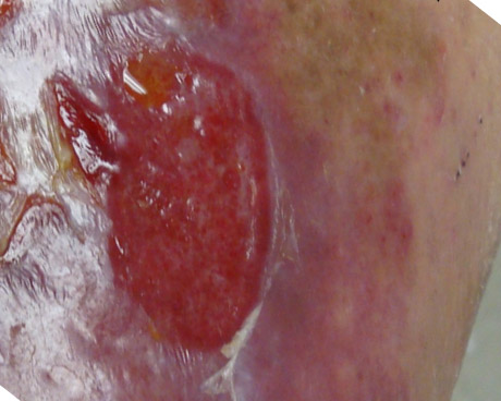
Earlier this week I was showing a patient that, while his wound might look like it was still open, in fact, most of it was covered with skin. When the epithelial cells first migrate, they are translucent and you have to get the light at an angle to determine whether there is actually “skin” over the wound. As the epithelium matures, it is more difficult to see the pink blood vessels below, and the melanin pigment usually activates.

Here are two examples of this – the same wound from slightly different angles – demonstrating epithelium. You do NOT want to accidentally traumatize this new skin. (And you can tell the wounds are nearly closed by the amount of drainage they have – which should be pretty small.)
Here’s another example in which from one angle of the light the wound looks open, but when you get the light from a different angle you can see that there’s skin over the wound:


Dr. Fife is a world renowned wound care physician dedicated to improving patient outcomes through quality driven care. Please visit my blog at CarolineFifeMD.com and my Youtube channel at https://www.youtube.com/c/carolinefifemd/videos
The opinions, comments, and content expressed or implied in my statements are solely my own and do not necessarily reflect the position or views of Intellicure or any of the boards on which I serve.


Dr. Fife,
Thank you for this article. Let me get your opinion on my experience with this “new thin epithelialization”. I have seen this many times and under ideal conditions this new thin epithelialization will mature over several weeks if the patient is very diligent with compression for a VLU and offloading for a DFU. Once they are discharged from our wound clinic it is uncertain how diligent they will be. What I frequently do is debride the wound including the new epithelialization up to the edges to allow the edges to migrate towards the center of the wound. This gives a stronger closure which I would feel much more confident with upon discharging a patient from the center. The edges won’t migrate if you leave the new epithelialization alone.
A great trick. ZnO2 cream also works to identify epithelium when the lighting is poor or you need to measure the wound dimensions , a neat trick based on simple magnetism properties. The skin is negatively charged so positively charged ZnO2 cream will stick, however when there is a loss of the epithelium, then the positively charged soft tissues will “repel” the skin cream. SImply measure from “white to white” and you have your dimensions of what is open.