

About six months ago I spent two days with some of my favorite people doing real time lymphatic imaging. The results have now been published as Case Report: The effect of automated manual lymphatic drainage therapy on lymphatic contractility in 4 distinct cases. Real-time lymphatic imagining was developed by Dr. Eva Sevick and her team including Drs. Melissa Aldrich and John Rasmussen. In addition to Dr. Sevick’s team at the Brown Foundation Institute of Molecular Medicine at The University of Texas Health Science Center at Houston, collaborators for this project included Irene Waldridge, Frank Aviles, and Drs. Ron Karni, Mark Melin, and me. We were evaluating the response to a novel device which performs automated manual lymphatic drainage therapy (AMLDT). Since this is my blog, I can tell you that the device is the Neuroglide, which is a pneumatic mat with 16 pressurized air channels that inflate and deflate to mimic the stretch and release action of manual lymphatic drainage therapy. The patient lies supine on the mat, placed over a somewhat firm surface.
The images in the small case series published in Frontiers in Medicine are interesting because almost no one has paid attention to the anatomy and function of the lymphatics along the spine. I have personally imaged hundreds of patients, but had never injected anyone’s back before. The inventor of the Neuroglide is the lovely Irene Waldridge, who is a household name in the industry because she founded Tactile Medical after developing the Flexitouch – a novel device for lymphedema management which was invented on her kitchen table. In 2021, her Neuroglide Back/Neck Pad, was FDA-cleared for sale to consumers. The Neuroglide is focused on the management of chronic pain by reducing inflammatory mediators through lymphatic stimulation. Her current company, Eva Medtec, is headquartered in Shakopee, MN, where devices are currently assembled in-house. Eva Medtec sponsored the project.
In one photo, Dr. Mark Melin (on right) is practicing his skill at intradermal injections on me. To obtain real time lymphatic images, micro doses of indocyanine green (ICG) are injected intradermally. Irene, wearing a blue sweater, is smiling because, for the first time in a long career of treating the lymphatics, was seeing the lymphatics work in real time.


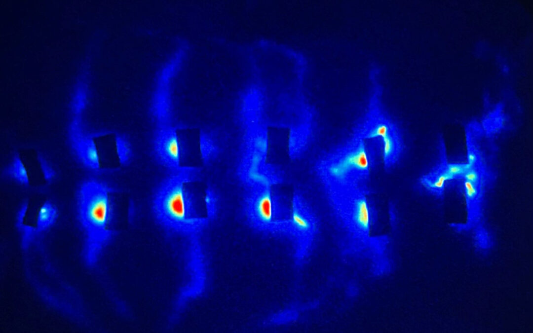
The other two images show the back of a subject lying completely prone, with the head to the right of the image and legs to the left. ICG has been injected at about every second or third vertebral level starting at the base of the neck. Those blue “bat wings” are the lymphatic fluid moving laterally from each injection site, around the flanks. The next image is the same subject lying on the left side, head to the right and legs to the left, so that you can see the lymphatics begin their rapid trip along the right lateral chest and flank – with some lymphatic fluid headed to the axillary lymphatics and others to the inguinal lymphatics. No one has ever seen images like this.


The paper, just published in Frontiers in Medical Technology, is now available Open Access (free). Be sure to download the movies and watch them!
–Caroline
(These photos are the property of Caroline Fife, MD and cannot be used without permission. The opinions here are mine and do not reflect those of Eva Medtec, with which I have no financial relationship.)

Dr. Fife is a world renowned wound care physician dedicated to improving patient outcomes through quality driven care. Please visit my blog at CarolineFifeMD.com and my Youtube channel at https://www.youtube.com/c/carolinefifemd/videos
The opinions, comments, and content expressed or implied in my statements are solely my own and do not necessarily reflect the position or views of Intellicure or any of the boards on which I serve.
