The Mysterious Case of the Ischemic Achilles
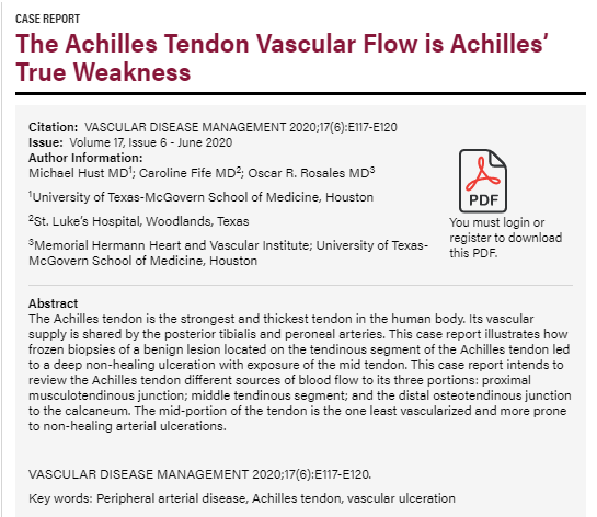
There’s a case report published online in “Vascular Disease Management” that is worth some conversation. The patient underwent a dermatological procedure on the back of the left calf that involved cryotherapy. About two weeks later, a wound opened several centimeters distal to the cryosurgery which exposed her Achilles tendon. She was seen by an excellent venous interventionalist who did a venous ablation that made no difference. She was seen at a wound center where the doctor confirmed that even though she was in her 80’s, she had no arterial disease according to an arterial Doppler. The lesion continued to get worse despite a variety of dressings. I was called because pyoderma gangrenosum (PG) was on the list of differential diagnoses and I seem to have a clinic full of patients with PG.
As you can see in the first photo, this lesion is not likely to be due to PG since there’s little evidence of any type of cellular response. It LOOKS ischemic which should be the first diagnosis considered anytime you are staring at a tendon. So, despite the fact her arterial Dopplers showed no evidence of arterial disease, I referred her to Dr. Oscar Rosales for an angiogram. He obtained a digital subtraction arteriogram of the left lower extremity which was significant for what he DIDN’T find. Just as her arterial Doppler had indicated, she had patent popliteal, posterior tibial, peroneal and anterior tibial arteries. But, the Achilles tendon is most commonly supplied by two arteries: the post tibial artery supplies the medial aspect of the Achilles and the peroneal supplies the lateral aspect via small branches that exist only for this purpose. As it happens, the Achilles branch of the peroneal artery was simply NOT THERE (see the link to this photo in the case report). The only explanation we have is that the cryosurgical procedure performed on the back of her calf superior to the wound destroyed this small artery and as the lateral Achilles became ischemic, a wound opened up from the inside.
(By the way, could everyone note the fact that if you take out even a small artery, the angiosome supplied by that vessel dies, and you get an “inside to outside” ulceration that is open down to the tissue level where you lost arterial supply – which is exactly what a Stage 4 pressure ulcer looks like – but I digress . . .)
Transdermal Oxygen to Facilitate the Development of Collateral Vessels
While we had found the cause of the problem – there wasn’t an obvious SOLUTION. Oscar Rosales is one of the finest invasive cardiologists in the country, but there was nothing he could do about an artery that was GONE. I decided to try something that I really didn’t think would work and that’s transdermal oxygen via the Oxyband dressing.
Approximately 12 weeks elapsed between the first photo showing the ischemic Achilles and the fifth photo showing a fully granulated wound. You can see that the undamaged posterior tibial artery is sending out collateral vessels to supply the lateral Achilles. You can actually SEE the blood vessels migrating from the medial to the lateral side of the Achilles and coming up through the Achilles, until the entire area is covered with granulation tissue. We stopped the Oxyband at this point and started using the dressings one would expect (collagen, etc.) It was another 12 weeks before there was epithelialization over the granulated wound, and that was facilitated by a single application of Theraskin.
So let’s review:
- Lower extremity ulcers that expose deep structures like tendons should undergo angiography unless there is a really obvious alternative explanation, because ischemia is always the most logical explanation for the exposure of deep structures.
- This is a classic case of an infarction of an angiosome (a three dimensional block of tissue which extends from bone or tendon to the skin and which is supplied by a vessel with a name). Angiosomal infarctions result in an ulceration that develops from the inside to the outside as the tissue layers necrose from the point of vascular occlusion to the terminus of the artery which is usually the skin (yes, just like severe pressure ulcers).
- An ulcer that exposes tendon is not VENOUS in origin, even if the patient also has venous insufficiency.
- Transdermal oxygen (via the Oxyband) seems to have facilitated the development of collateral vessels.

Dr. Fife is a world renowned wound care physician dedicated to improving patient outcomes through quality driven care. Please visit my blog at CarolineFifeMD.com and my Youtube channel at https://www.youtube.com/c/carolinefifemd/videos
The opinions, comments, and content expressed or implied in my statements are solely my own and do not necessarily reflect the position or views of Intellicure or any of the boards on which I serve.


