

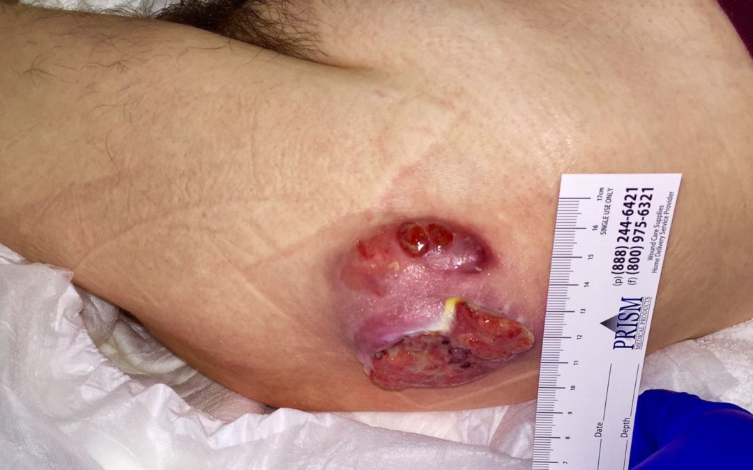
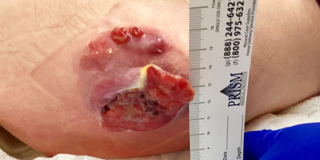
An adult patient with severe cerebral palsy developed several pressure ulcers after hospital admission for pneumonia. This one has persisted for many months. While it’s possible that the trauma that initiated it was a pressure injury, this is pyoderma gangrenosum. Note the ruffled, hypertrophic border, the purple margin and the new breakdown with hypertrophic vascular tissue along the edge. This was a very difficult case to treat and never fully resolved – although it did improve with oral steroids, intralesional Kenalog, oral cyclosporin and a variety of other interventions.
My goal with this case is not to discuss the treatment of PG (which is always challenging), but to show some of its many faces.
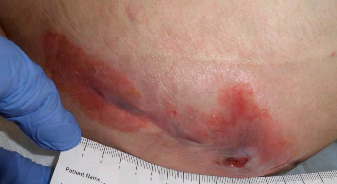
This is the best we managed to get it under control.
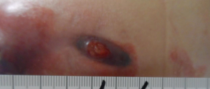
Although this is a blurry photo, this is how the PG looks as it activates again – easily mistaken for pressure but that’s the characteristic purple margin.

Dr. Fife is a world renowned wound care physician dedicated to improving patient outcomes through quality driven care. Please visit my blog at CarolineFifeMD.com and my Youtube channel at https://www.youtube.com/c/carolinefifemd/videos
The opinions, comments, and content expressed or implied in my statements are solely my own and do not necessarily reflect the position or views of Intellicure or any of the boards on which I serve.
