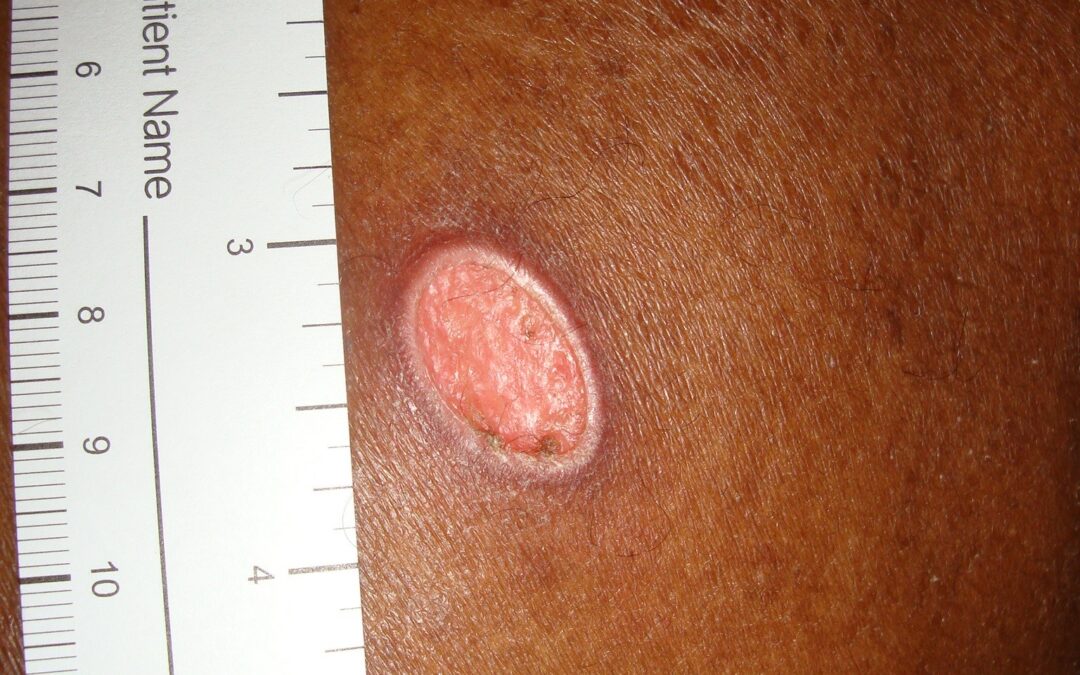
Wang and colleagues recently published an article in JAMA Dermatology about primary cutaneous cryptococcus. The article is not available open access, so we can’t look at the photos of the case. However, in 2012, Dr. Adelaide Hebert and I published a case report of cutaneous cryptococcosis in the Journal of the American Academy of Dermatology, so I thought I’d post a photo of my case from a decade ago. This wound looks like no big deal. In fact, it looks like it has skin over it, doesn’t it? Unrecognized cryptococcosis on the upper back of a 64-year-old patient with an acquired ichthyosis on chronic immunosuppression caused by renal transplantation.
The patient was a 64-year-old African American man referred to me by the transplant team. He told me that the problem started after he scratched his itchy back against a tree. At the time of the contact with the tree bark, he told me that he was fully clothed including a shirt and thin jacket. At the time of my initial evaluation, I thought he was healing, although the skin that seemed to cover the area had no melanin. The lesion measured 1.5 × 1.4 × 0.2 cm. What did not make sense was that he complained that his pain was 7 on a scale of 10. I put a hydrocolloid dressing over it hoping that would help the pain, but after a month of conservative care, it was no better. So, I did a biopsy.
The biopsy showed numerous eosinophilic microorganisms located both within histiocytes and free within the tissues. Periodic acid–Schiff (PAS) staining highlighted fungal organisms that were present. Sadly, back in 2012 I didn’t have next generation DNA sequencing which saved the life of a different transplant patient with mucormycosis. As soon as the biopsy results were available, I alerted the transplant team but by then the patient had developed shortness of breath and was admitted to the hospital with pneumonia and did not survive. Cultures of tissue (eventually) grew Cryptococcus.
The Wang article made me remember this long-ago patient – and how innocuous the lesion looked. We have better technology for diagnosing rare infections now – the patient might have survived if I had managed to get a diagnosis earlier. I thought I would post this as a reminder to biopsy patients with immune suppression.

Dr. Fife is a world renowned wound care physician dedicated to improving patient outcomes through quality driven care. Please visit my blog at CarolineFifeMD.com and my Youtube channel at https://www.youtube.com/c/carolinefifemd/videos
The opinions, comments, and content expressed or implied in my statements are solely my own and do not necessarily reflect the position or views of Intellicure or any of the boards on which I serve.


