

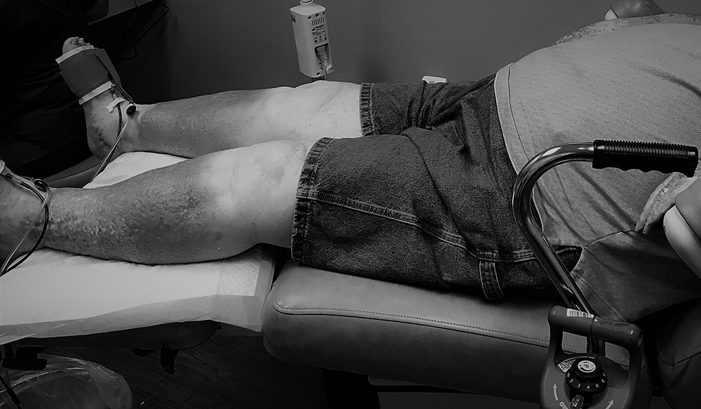
This is an 80 year old man with severe bilateral lower extremity lymphedema. I hospitalized him the day I took these photos. He had copious amounts of neon green drainage and increasing erythema of his legs, but what really precipitated hospital admission was his increasing shortness of breath. He couldn’t sleep unless sitting bolt upright using 4 pillows and his saturation on room air was 84%.
He is obese with type 2 diabetes for 8 years, COPD (on oxygen for 10 years), congestive heart failure (CHF), atrial fibrillation, and sleep apnea. The one thing he has going for him is that when I performed arterial screening on his first visit (per the US Wound Registry arterial screening quality measure criteria), his peripheral arterial supply was good, based on skin perfusion pressure and pulse volume recording.
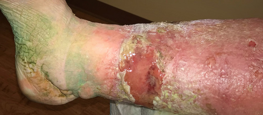
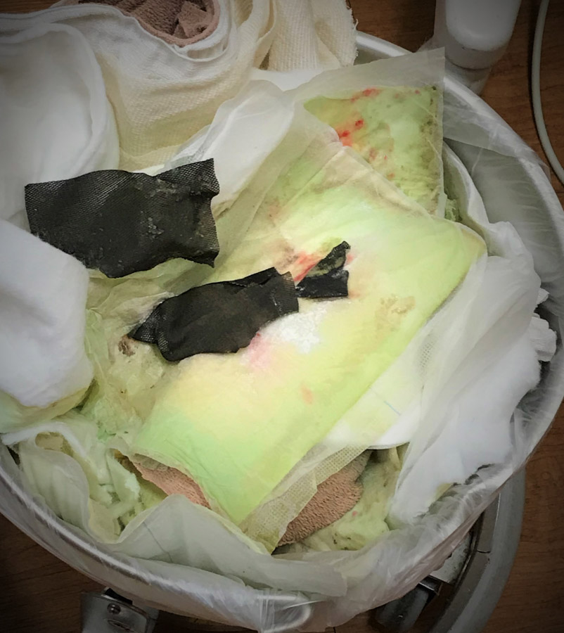
Because his arterial status was adequate, I had applied aggressive compression to both legs. Unfortunately, at his next visit, the day I took these photos, he was dramatically worse. The green staining of his skin is most likely from Pseudomonas colonization. These huge, new open blisters actually developed UNDER compression wraps because he is profoundly volume overloaded. Although he clearly has venous insufficiency (evidenced by hemosiderin and lipodermatosclerosis), the huge blisters are actually caused by his skin splitting apart from his terrible leg edema, which is caused by his heart failure. Although I suppose it’s not disingenuous to call these venous leg ulcers (and probably most clinicians do), these lesions are really more accurately classified as chronic ulcers due to his underlying medical conditions.

I have been told by some venous interventional colleagues that patients like this can be managed with venous ablation and compression and that venous ablation should be the lynchpin of treatment. However, he’s already had several venous ablation procedures and even though I optimized his compression with 4 layer wraps, I couldn’t control his edema. In fact, I have a clinic full of heart failure patients with volume overload and orthopnea who are chair sleepers. I can’t get control of their leg edema (which extends to the abdomen) with typical venous compression wraps that stop below the knee. Patients like this need better management of their heart failure. Additionally, it’s almost impossible for venous wraps to overcome the intra-abdominal pressure caused by an obese abdomen and COPD. This is a multi-factorial problem.
This patient has one of the chronic condition “triads” that Medicare fears most. Triads are groupings of specific diagnoses which, when present together, tell Medicare they will spend at least $60,000 per year on treatment and the patient will need more than 20 outpatient visits annually. The specific diagnoses are: CHF, a fib, COPD/asthma, chronic renal disease, stroke and depression. This particular patient’s triad is: CHF, A fib and COPD. I guarantee he’s already made more than 20 outpatient visits this year, and that $60,000 is an underestimation of his 2018 healthcare costs.
In a future post I will explain why patients like this represent the “Bermuda triad” for outpatient wound care when payers and providers attempt to create “episodes of care” for wounds caused by chronic disease.

Dr. Fife is a world renowned wound care physician dedicated to improving patient outcomes through quality driven care. Please visit my blog at CarolineFifeMD.com and my Youtube channel at https://www.youtube.com/c/carolinefifemd/videos
The opinions, comments, and content expressed or implied in my statements are solely my own and do not necessarily reflect the position or views of Intellicure or any of the boards on which I serve.
