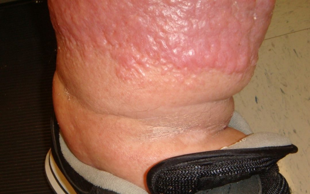
This just out in the journal Obesity –– an article I co-authored on Lymphatic Function and Anatomy in Early Stages of Lipedema (John C. Rasmussen, Melissa B. Aldrich, Caroline E. Fife, Karen L. Herbst, Eva M. Sevick-Muraca; 15 June 2022).
Lipedema is a genetically mediated pathological accumulation of fat, usually below the waist and in women, that is diet resistant. It’s considered an inflammatory subcutaneous adipose tissue disease that may progress to lipolymphedema, a condition similar to lymphedema. The question we wanted to answer was whether the lymphatics of patients with early lipedema was the same or different from the lymphatic abnormalities we have identified in patients with lymphedema. In patients with lymphedema related to cancer and in primary lymphedema, real time lymphatic imaging with indocyanine green shows dilated lymphatic vessels, impaired pumping, and dermal backflow, so we know what abnormal lymphatics look like – thanks to the novel real-time lymphatic imaging developed by Dr. Eva Sevick.
We did a pilot study of 20 women with Stage 1 or 2 lipedema that had not progressed to lipolymphedema, and assessed their upper and lower extremities using near-infrared fluorescence lymphatic imaging. We compared them to a control population of similar age and BMI. The study showed that in early lipedema sufferers, lower extremity lymphatic vessels were dilated and showed intravascular pooling, but the propulsion rates of the lymphatics significantly exceeded those of control individuals. (The upper extremity lymphatics of individuals with lipedema were unremarkable.) In contrast to individuals with lymphedema, individuals with Stage I and II lipedema did not exhibit dermal backflow.
In other words, the lymphatics of early lipedema patients appear quite different from those of lymphedema patients. Lipedema patients have lymphatic vessel dilation but don’t have lymphatic dysfunction – at least, early in the lipedema disease process. Something definitely goes wrong later (at least in some patients) to cause lipolymphedema, but the process is somehow different than lymphedema.
Here are some previous blog articles I have posted on lymphedema, lipedema and/or IC green imaging:
- Lymphatic Dysfunction in Cancer Patients – Insights Into This Mysterious and Vital Organ System
- What the Heck is That Thing? Massive Localized Lymphedema Talk Now Available
- Axillary Web Syndrome Revealed
- New Study Shows That Even Patients With Early Venous Disease Have a Degradation in Lymphatic Function. Use My Personal Share Link to Obtain a Free Copy!
- Why My Blue Mondays are Happy
- If Success and Failure Look the Same Clinically, We Have a Problem
- The Wound Whisperer – Lymphedema of the Plantar Foot
- 3 Patients with Lymphedema of the Hand – One of Them Has Open Wounds – Why?
- What is it Woundsday
- Lipedema & Lymphedema Patient Round Table Discussions Available on YouTube
- Wounds Are a Symptom: If Only the Surgeon Had Noticed Her Lymphedema
- The Lymphedema Therapist: A Wound Physician’s Secret Weapon?
- Visualizing the Relationship Between Venous Insufficiency and Lymphedema
- The Inconvenient Truth in a Convenience Sample – the Prevalence of Lymphedema in a Wound Clinic
- In case you missed it in 2017: A Taxonomy For Skin Disorders in Lymphedema Patients

Dr. Fife is a world renowned wound care physician dedicated to improving patient outcomes through quality driven care. Please visit my blog at CarolineFifeMD.com and my Youtube channel at https://www.youtube.com/c/carolinefifemd/videos


